Text: Nienke Beintema
New video footage made by Wageningen researchers reveal a fascinating sight. It looks as though elongated tadpoles are tumbling and swirling around in a network of fast-flowing streams. Only these ‘tadpoles’ are actually trypanosomes and the ‘streams’ are the blood vessels of zebrafish.
Trypanosomes are parasites that cause sleeping sickness in Africa and Chagas disease in Latin America (see text box). In the footage they seem to be float through the blood vessels in an aimless, almost lazy fashion. They don’t use their flagellum – or ‘tail’ – for propulsion, as has always been presumed, but go wherever the bloodstream takes them. They do, however, stick to the wall of a blood vessel with their backs and then enter their host’s tissue. This is important new information, offering a starting point in the battle against these diseases. The results were published in the September issue of the scientific journal eLife.
Neglected diseases
‘Sleeping sickness and Chagas disease are what we call neglected diseases,’ says Maria Forlenza, associate professor at the Cell Biology and Immunology group. ‘Worldwide they cause hundreds of thousands of deaths per year, but they receive relatively little attention. Why? Because they are less deadly than diseases like malaria. And because it is very difficult to find funding for the kind of fundamental research that is needed to understand how these diseases work.’
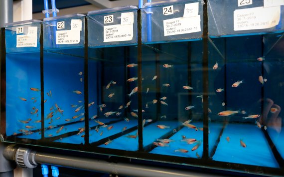
Forlenza and her colleagues have revealed for the first time how the parasite moves and behaves in a host. ‘We couldn’t use conventional laboratory animals like mice because you can’t look into their moving bloodstreams through a microscope. That’s why we used live zebrafish. Very young zebrafish are transparent so you can see what is happening in their moving bloodstream.’
In these in vivo studies, the researchers were able to prove that current models and assumptions about how trypanosomes move and infect their hosts, based on in vitro research and computer models, are incorrect. Forlenza: ‘To me, that is one of the most important messages of our study: in some cases, you still need live hosts. Nowadays there is a strong trend towards reducing animal experiments, which I fully support – but we have shown that it sometimes has added value.’
Clinging
It was previously thought that trypanosomes use their ‘tail’ to move around actively in the bloodstream. Hence, strategies to block this type of parasite have focused on the motor proteins that make their tails move. The footage that Forlenza and her colleagues filmed at 500 frames per second shows that there is no active propulsion.
The footage also reveals how the parasite attaches itself to the walls of blood vessels. ‘This attachment is very similar to that of our own immune cells’, explains Forlenza. ‘Just like white blood cells, the parasites can attach themselves to the walls of veins, but not to those of arteries. Apparently there is something in the make-up of vein walls that makes them penetrable – and the parasite has found a way of moving in and out of the bloodstream just like our own immune cells.’
Glue protein
The footage showed that the parasite first touches the wall of the vein with its back. Interestingly enough, this contact site is not on the same side as the tail. Forlenza: ‘While the parasite is attached to the vein wall, the flagellum is still free to move. It may play a role in pushing the parasite through the wall, into the tissue.’
The researchers suspect that, just like immune cells, trypanosomes use adhesion molecules, or ‘glue’ molecules, on their surface to attach themselves to proteins on the surface of vein cells. ‘We are now zooming in on which proteins these may be’, says Forlenza, ‘and we have found some very promising candidates.’
These diseases cost thousands of lives every year but get very little attention
What if you could block this glue protein, either with antibodies or by blocking the gene that codes for the glue protein? Forlenza and colleagues explored this possibility by producing mutant trypanosomes in which they labelled different surface proteins with colours that are distinguishable under the microscope. ‘One of the mutant proteins seems to be located exactly where the parasite attaches to the host’, says the researcher. ‘Now we have a nice starting point for our follow-up research: to see what happens if you alter or disable this protein in the parasite – or if you create antibodies that target it. I am pretty sure that one of these routes will prove successful in the fight against these diseases.
Seeing is believing
Forlenza is currently writing grant proposals for this follow-up research, together with the University of Cambridge. ‘In the UK there is more funding for fundamental research on neglected diseases.’ This research also requires collaboration within the WUR Department of Animal Sciences. ‘My own group, Cell Biology and Immunology, contributes in the area of host-pathogen interactions’, says Forlenza, ‘and the biomechanics expertise comes from the Experimental Zoology group.’
It may take at least 10 to 15 years before this research results in a clinical application such as a vaccine against sleeping sickness. But there are no shortcuts, says Forlenza. ‘You really need this fundamental knowledge. For years we worked on the basis of assumptions about trypanosomes, but what you need is in vivo evidence. ‘Seeing is believing’, is our motto.’
ALMOST ALWAYS FATAL
Trypanosomes are unicellular parasties that cause sleeping sickness in Africa and Chagas disease in South America. Sleeping sickness is transmitted by the tsetse fly, while Chagas is spread by beetle-like biting bugs. The first symptoms are fever, headache, itching and aching joints. In a later stage, neurological symptoms occur, such as disturbed sleep, confusion and memory loss. Coma and death almost always follow. Sleeping sickness kills around 10,000 people in Africa every year, although some sources cite numbers between 50,000 and 500,000. In South America, there are thought to be about 8000 deaths a year.

 Photo: Aldo Allessi