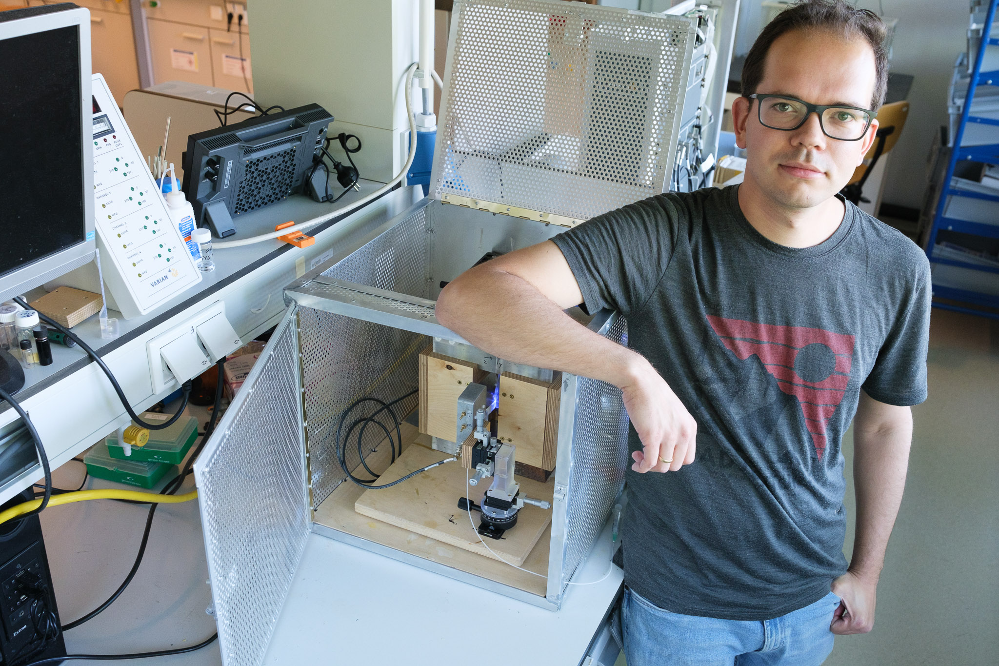
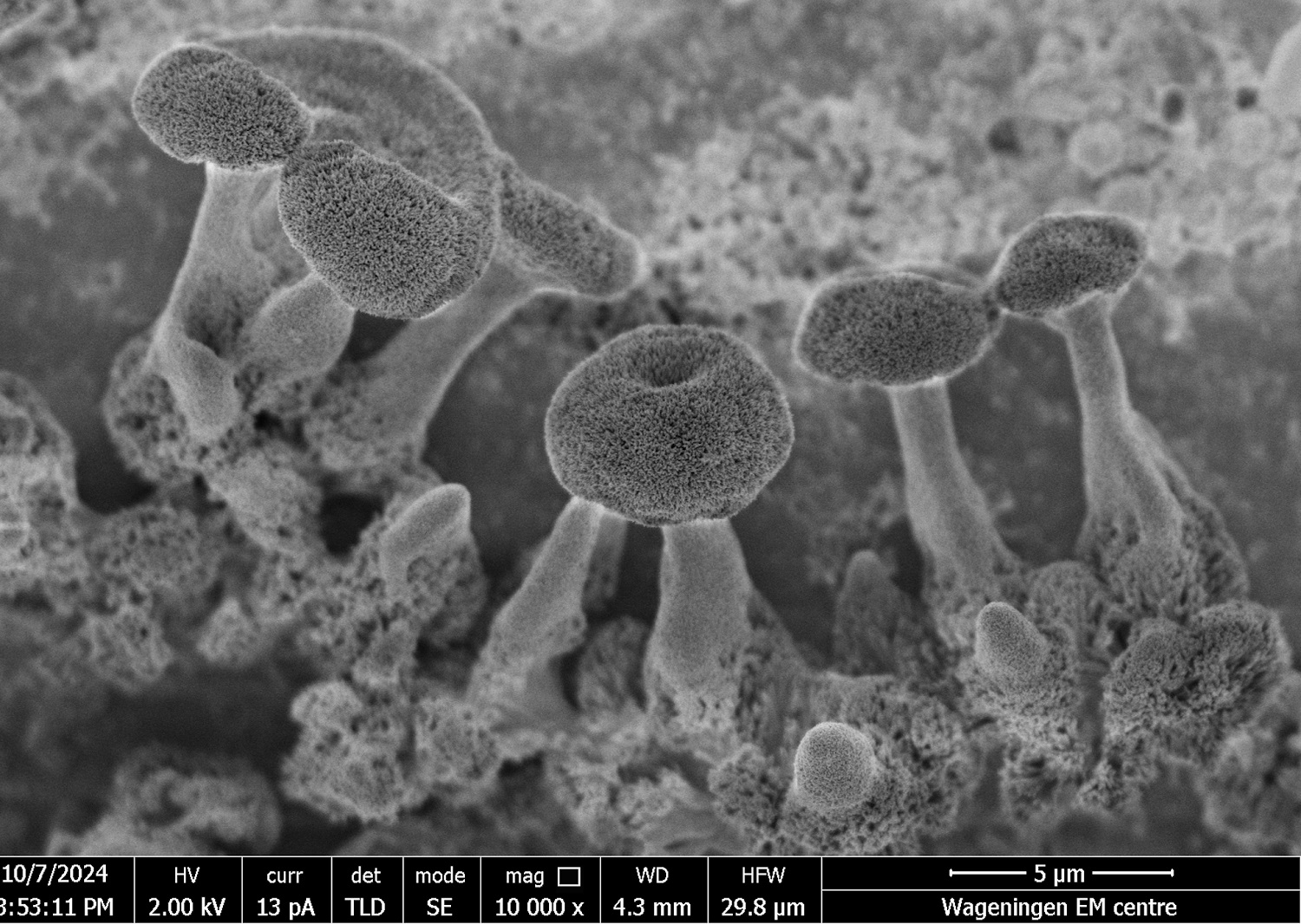
 Photo Vittorio Saggiomo, Wageningen Electron Microscopy Centre
 No, this is not a black-and-white photo of a cluster of sulphur tuft fungi. While they look real, they are in fact sand and salt crystals. A group of students made the mini artwork during the BioNanoTechnology Introduction practical classes. With a big dose of luck, teacher Anton Bunschoten happily admits. ‘It was a lucky shot.’
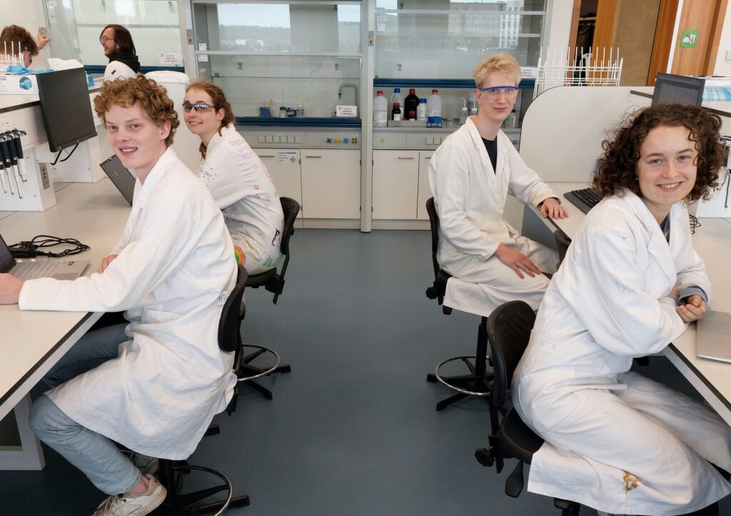
But that doesn’t make the result any less impressive. The picture vies with the crystals that got Harvard scientist Nadir Kaplan and his team a publication in Science in 2017. And that is why Bunschoten is proud of what Joppe van Eck, Sophie Hennink, Aron van de Wetering and Tara van Veen achieved in a couple of afternoons.
Gold particles
In the practical taught by Bunschoten, students learn how to make nanoparticles. ‘In the first four weeks, we specify the experiments. For example, we get them to make gold nanoparticles of the kind used in diagnostic tests such as the Covid test. The two coloured lines in that test consist of such gold nanoparticles. In the final two weeks, they can make up their own experiments.’
The fungi group decided to attempt a rerun of Kaplan’s work. That basically meant crystalizing solutions of silicates (sand) and barium chloride (salt) from the air under the influence of CO₂. Bunschoten: ‘Depending on the acidity and the supply of CO₂, the crystals will grow into a certain shape. Kaplan’s team got plants and trees; they were in full control of the outcome.’
Bingo
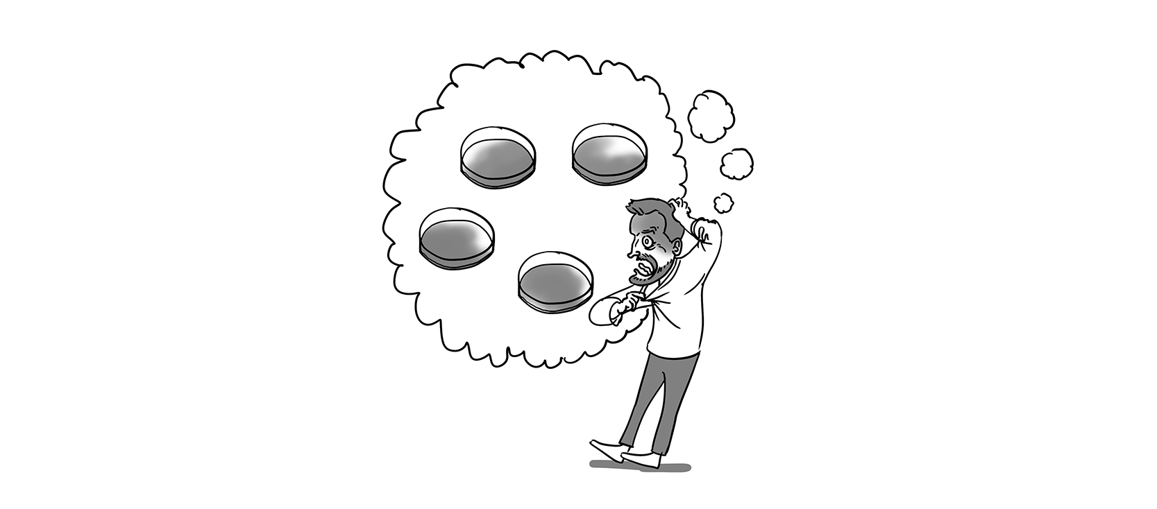
Three of the students’ four attempts failed. But it was bingo for the fourth one, they discovered when the photos taken by post-doc Vittorio Saggiomo came back. Fungi can clearly be seen in the photos, which were taken using the equipment of the Wageningen Electron Microscopy Centre (WEMC) in Radix. According to Bunschoten, the reason is not clear. ‘Two completely different shapes arose, which suggests two growth phases.’
Jellyfish
The fungi are only visible if you magnify the whitish stuff by a factor of 10,000. Under an ordinary light microscope it looks more like a swarm of jellyfish, as Tara van Veen shows on her laptop. The students have since tried to repeat the experiment. Whether that worked will become clear next week when Saggiomo looks at the result under the electron microscope.